Skin cancer treatment and reconstruction

Skin Cancer: Symptoms, Types, Diagnosis, and Treatment
Skin cancer is the most common type of cancer worldwide. It develops when skin cells grow abnormally, usually due to damage from ultraviolet (UV) radiation from the sun or tanning beds. When detected early, most forms of skin cancer can be treated effectively, often with simple surgical procedures. However, if left untreated, some types can spread and become life-threatening.
Causes and Risk Factors
Skin cancer is most often caused the following risk factors:
Fair skin, light eyes, and hair color.
Frequent sunburns, especially in childhood.
Use of tanning beds.
Family history of skin cancer.
Weakened immune system (HIV, organ transplant, long-term medications).
Older age (but melanoma is also common in younger adults).
Chronic scars or burns.
-
The most common form of skin cancer (about 80% of cases).
Slow-growing and rarely spreads to other parts of the body.
Signs include: a shiny bump, visible blood vessels, or a sore that heals and reopens.
-
More aggressive than BCC and may spread if untreated.
Often appears as red, scaly patches, non-healing ulcers, or wart-like lesions.
Can develop in areas of long-standing scars, wounds, or burns.
-
The most serious type of skin cancer.
Can spread quickly to other organs if not treated early.
Usually presents as an irregular mole or dark spot that changes in size, shape, or color.
-
One half doesn’t match the other.
-
Uneven, irregular edges.
-
Multiple colors (brown, black, red, blue).
-
Larger than 6 mm (pencil eraser size)
-
Any change in appearance over time
Signs and Symptoms
See a dermatologist/plastic surgeon if you notice:
A new growth or sore that does not heal within 3 weeks.
Changes in an existing mole (color, size, shape).
Itching, bleeding, or tenderness in a mole or spot.
Rough, scaly patches or ulcers that don’t go away.
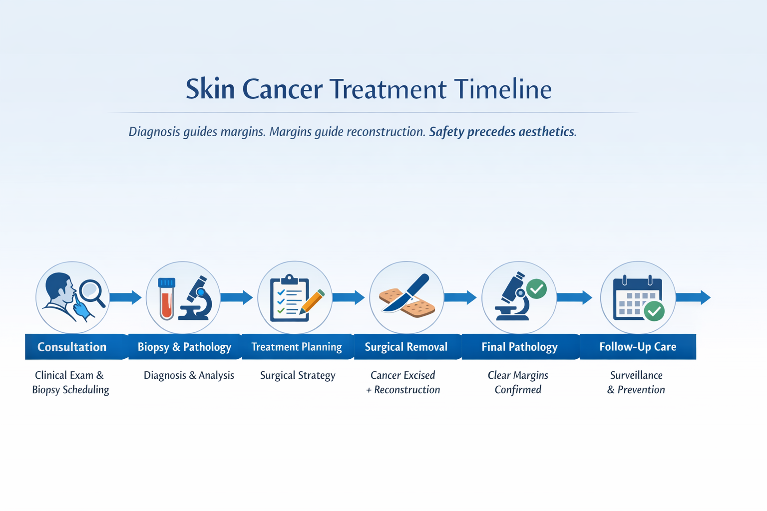
Diagnosis: How Skin Cancer Is Detected
Diagnosis is usually made with a skin biopsy:
Excisional biopsy – the entire lesion is removed (preferred for pigmented lesions).
Incisional biopsy – a part of the lesion is removed (for large or complex cases or areas of cosmetic concern like eyes, ears and nose).
The pathologist then examines the tissue under a microscope to confirm if it is cancer, identify the type of tumor, and assess its risk features. These findings guide the next steps, whether the lesion should be excised with wider margins or whether no further treatment is needed.
Skin Cancer Treatment Options
Treatment depends on the type, size, and stage of cancer.
Surgical removal (excision): The most effective treatment, with margins starting around 3 mm depending on cancer type. Reconstruction may involve sutures, skin flaps, or grafts. Surgery has the lowest recurrence risk (~3–5%). Mohs is the gold standard.
Cryotherapy: Freezing the lesion with liquid nitrogen, mainly for early or precancerous spots.
Radiation therapy: Used when surgery is not possible or after surgery when certain types of skin cancer exhibit high risk factors.
Chemotherapy / Immunotherapy: For advanced or metastatic melanoma/SCC.
In all cases, clear surgical margins are confirmed before reconstruction, especially for melanoma.
Reconstruction After Skin Cancer Surgery
Once a skin cancer has been completely removed, the next step is often reconstruction. The goal of reconstruction is not only to close the wound but also to restore both function and natural appearance.
Primary closure (stitches): For small lesions, the wound can usually be closed directly with sutures, leaving a fine scar.
Skin flaps: For larger or more complex wounds, surrounding tissue can be carefully rotated, advanced, or repositioned to cover the defect. Flaps use the patient’s own skin, which blends best in terms of texture and color.
Skin grafts: In some cases, skin is taken from another area of the body and transplanted to cover the surgical site. This is common when there isn’t enough local tissue to close the wound.
Specialized techniques: For sensitive areas such as the nose, lips, eyelids, or ears, advanced reconstructive methods (combining all of the above) are used to maintain normal function while minimizing visible scarring.
Reconstruction is always planned with a balance between oncologic safety (making sure all cancer is removed with clear margins) and aesthetic outcome.


A basal cell carcinoma on the nose reconstructed with a bilobed flap. This will fade in time.
Frequently asked questions in my practice
-
In most cases, skin cancers grow locally and do not spread rapidly over short periods. Once diagnosed, surgery is scheduled appropriately based on urgency and pathology type.
Treatment is planned methodically,
not rushed, not delayed unnecessarily.
-
If final pathology shows that cancer cells extend to the edge of the specimen, additional excision is performed to ensure complete removal.
Clear margins are confirmed through pathology, not visual inspection alone. They are the single most important factor of recurrence-free disease.
-
Any surgical removal creates a scar. The goal is to place incisions strategically and reconstruct in a way that restores contour and function.
Scar appearance improves over time, and scar care guidance is provided during follow-up.
Oncologic safety comes first. Aesthetic planning follows.
-
In certain cases, particularly melanoma or high-risk tumors, additional procedures or imaging may be required.
Most skin cancers are treated definitively in a single surgical session. If further steps are needed, they are discussed clearly and promptly. In some cases, aesthetic refinement in a second or third surgery is needed to complete the result.
-
Follow-up frequency depends on the cancer type and risk profile. Some patients require periodic skin checks, while others may need structured surveillance.
Monitoring is part of treatment, not an afterthought. It is scheduled ahead of time with the dermatologist/oncologist.